General Dermatology
We offer skin cancer screening. The added benefit is that we are likely able to manage the skin cancer in the same facility that it was diagnosed with our team of experts. Not every skin cancer needs surgery. We are able to address all of your concerns and options for an individualized treatment plan.
Skin Cancer Screening
Skin cancer is the most common form of human cancers, affecting more than one million Americans every year. One in five Americans will develop skin cancer at some point in their lives. Skin cancers are generally curable if caught early. However, people who have had skin cancer are at a higher risk of developing a new skin cancer, which is why regular self-examination and doctor visits are imperative.
The vast majority of skin cancers are composed of three different types: basal cell carcinoma, squamous cell carcinoma and melanoma.
Basal Cell Carcinoma
This is the most common form of skin cancer. Basal cells reside in the deepest layer of the epidermis, along with hair follicles and sweat ducts. When a person is overexposed to UVB radiation, it damages the body’s natural repair system, which causes basal cell carcinomas to grow. These tend to be slow-growing tumors and rarely metastasize (spread). Basal cell carcinomas can present in a number of different ways:
- raised pink or pearly white bump with a pearly edge and small, visible blood vessels
- pigmented bumps that look like moles with a pearly edge
- a sore that continuously heals and re-opens
- flat scaly scar with a waxy appearance and blurred edges
Despite the different appearances of the cancer, they all tend to bleed with little or no cause. Eighty-five percent of basal cell carcinomas occur on the face and neck since these are areas that are most exposed to the sun.
Risk factors for basal cell carcinoma include having fair skin, sun exposure, age (most skin cancers occur after age 50), exposure to ultraviolet radiation (as in tanning beds) and therapeutic radiation given to treat an unrelated health issue.
Diagnosing basal cell carcinoma requires a biopsy — either excisional, where the entire tumor is removed along with some of the surrounding tissue, or incisional, where only a part of the tumor is removed (used primarily for large lesions).
Treatments for basal cell carcinoma include:
- Cryosurgery Some basal cell carcinomas respond to cryosurgery, where liquid nitrogen is used to freeze off the tumor.
- Curettage and Desiccation — The preferred method of dermatologists, this treatment involves using a small metal instrument (called a curette) to scrape out the tumor along with an application of an electric current into the tissue to kill off any remaining cancer cells.
- Mohs Micrographic Surgery — The preferred method for large tumors, Mohs Micrographic Surgery combines removal of cancerous tissue with microscopic review while the surgery takes place. By mapping the diseased tissue layer by layer, less healthy skin is damaged when removing the tumor.
- Prescription Medicated Creams — These creams can be applied at home. They stimulate the body’s natural immune system over the course of weeks.
- Radiation Therapy — Radiation therapy is used for difficult-to-treat tumors, either because of their location, severity or persistence.
- Surgical Excision — In this treatment the tumor is surgically removed and stitched up.
Squamous Cell Carcinoma
Squamous cells are found in the upper layer (the surface) of the epidermis. They look like fish scales under a microscope and present as a crusted or scaly patch of skin with an inflamed, red base. They are often tender to the touch. It is estimated that 250,000 new cases of squamous cell carcinoma are diagnosed annually, and that 2,500 of them result in death.
Squamous cell carcinoma can develop anywhere, including inside the mouth and on the genitalia. It most frequently appears on the scalp, face, ears and back of hands. Squamous cell carcinoma tends to develop among fair-skinned, middle-aged and elderly people who have a history of sun exposure. In some cases, it evolves from actinic keratoses, dry scaly lesions that can be flesh-colored, reddish-brown or yellow black, and which appear on skin that is rough or leathery. Actinic keratoses spots are considered to be precancerous.
Like basal cell carcinoma, squamous cell carcinoma is diagnosed via a biopsy — either excisional, where the entire tumor is removed along with some of the surrounding tissue, or incisional, where only a part of the tumor is removed (used primarily for large lesions).
Treatments for squamous cell carcinoma include:
- Cryosurgery Some basal cell carcinomas respond to cryosurgery, where liquid nitrogen is used to freeze off the tumor.
- Curettage and Desiccation — The preferred method of dermatologists, this treatment involves using a small metal instrument (called a curette) to scrape out the tumor along with an application of an electric current into the tissue to kill off any remaining cancer cells.
- Mohs Micrographic Surgery — The preferred method for large tumors, Mohs Micrographic Surgery combines removal of cancerous tissue with microscopic review while the surgery takes place. By mapping the diseased tissue layer by layer, less healthy skin is damaged when removing the tumor.
- Prescription Medicated Creams — These creams can be applied at home. They stimulate the body’s natural immune system over the course of weeks.
- Radiation Therapy — Radiation therapy is used for difficult-to-treat tumors, either because of their location, severity or persistence.
- Surgical Excision — In this treatment the tumor is surgically removed and stitched up.
Melanoma
While melanoma is the least common type of skin cancer, it is by far the most virulent. It is the most common form of cancer among young adults age 25 to 29. Melanocytes are cells found in the bottom layer of the epidermis. These cells produce melanin, the substance responsible for skin pigmentation. That’s why melanomas often present as dark brown or black spots on the skin. Melanomas spread rapidly to internal organs and the lymph system, making them quite dangerous. Early detection is critical for curing this skin cancer.
Melanomas look like moles and often do grow inside existing moles. That’s why it is important for people to conduct regular self-examinations of their skin in order to detect any potential skin cancer early, when it is treatable. Most melanomas are caused by overexposure to the sun beginning in childhood. This cancer also runs in families.
Melanoma is diagnosed via a biopsy. Treatments include surgical removal, radiation therapy or chemotherapy.
What to Look For
The key to detecting skin cancers is to notice changes in your skin. Look for:
- Large brown spots with darker speckles located anywhere on the body.
- Dark lesions on the palms of the hands and soles of the feet, fingertips toes, mouth, nose or genitalia.
- Translucent pearly and dome-shaped growths.
- Existing moles that begin to grow, itch or bleed.
- Brown or black streaks under the nails.
- A sore that repeatedly heals and re-opens.
- Clusters of slow-growing scaly lesions that are pink or red.
The American Academy of Dermatology has developed the following ABCDE guide for assessing whether or not a mole or other lesion may be becoming cancerous.
Asymmetry: Half the mole does not match the other half in size, shape or color.
Border: The edges of the mole are irregular or blurred.
Color: The mole is not the same color throughout.
Diameter: The mole is larger than one-quarter inch in size.
Elevation: The mole becomes elevated or raised from the skin.
If any of these conditions occur, please make an appointment to see one of our dermatologists right away. The doctor may do a biopsy of the mole to determine if it is or isn’t cancerous.
Prevention
Roughly 90% of nonmelanoma cancers are attributable to ultraviolet radiation from the sun. That’s why prevention involves:
-
- Staying out of the sun during peak hours (between 10 a.m. and 4 p.m.).
- Covering up the arms and legs with protective clothing.
- Wearing a wide-brimmed hat and sunglasses.
- Using sunscreens year round with a SPF of 15 or greater and sunblocks that work on both UVA and UVB rays. Look for products that use the term “broad spectrum.”
- Checking your skin monthly and contacting your dermatologist if you notice any changes.
- Getting regular skin examinations. It is advised that adults over 40 get an annual exam with a dermatologist.
Acne/Acne Rosacea
Acne is the most frequent skin condition in the United States. It is characterized by pimples that appear on the face, back and chest. Every year, about 80% of adolescents have some form of acne and about 5% of adults experience acne.
Acne is made up of two types of blemishes:
- Whiteheads/Blackheads, also known as comedones, are non-inflammatory and appear more on the face and shoulders. As long as they remain uninfected, they are unlikely to lead to scarring.
- Red Pustules or Papules are inflamed pores that fill with pus. These can lead to scarring.
Causes
In normal skin, oil glands under the skin, known as sebaceous glands, produce an oily substance called sebum. The sebum moves from the bottom to the top of each hair follicle and then spills out onto the surface of the skin, taking with it sloughed-off skin cells. With acne, the structure through which the sebum flows gets plugged up. This blockage traps sebum and sloughed-off cells below the skin, preventing them from being released onto the skin’s surface. If the pore’s opening is fully blocked, this produces a whitehead. If the pore’s opening is open, this produces blackheads. When either a whitehead or blackhead becomes inflammed, they can become red pustules or papules.
It is important for patients not to pick or scratch at individual lesions because it can make them inflamed and can lead to long-term scarring.
Treatment
Treating acne is a relatively slow process; there is no overnight remedy. Some treatments include:
-
- Benzoyl Peroxide — Used in mild cases of acne, benzoyl peroxide reduces the blockages in the hair follicles.
- Oral and Topical Antibiotics — Used to treat any infection in the pores.
- Hormonal Treatments — Can be used for adult women with hormonally induced acne.
- Tretinoin — A derivative of Vitamin A, tretinoin helps unplug the blocked-up material in whiteheads/blackheads. It has become a mainstay in the treatment of acne.
- Extraction — Removal of whiteheads and blackheads using a small metal instrument that is centered on the comedone and pushed down, extruding the blocked pore.
Eczema
More information about our eczema treatments coming soon.
Psoriasis
Psoriasis is a skin condition that creates red patches of skin with white, flaky scales. It most commonly occurs on the elbows, knees and trunk, but can appear anywhere on the body. The first episode usually strikes between the ages of 15 and 35. It is a chronic condition that will then cycle through flare-ups and remissions throughout the rest of the patient’s life. Psoriasis affects as many as 7.5 million people in the United States. About 20,000 children under age 10 have been diagnosed with psoriasis.
In normal skin, skin cells live for about 28 days and then are shed from the outermost layer of the skin. With psoriasis, the immune system sends a faulty signal which speeds up the growth cycle of skin cells. Skin cells mature in a matter of 3 to 6 days. The pace is so rapid that the body is unable to shed the dead cells, and patches of raised red skin covered by scaly, white flakes form on the skin.
Psoriasis is a genetic disease (it runs in families), but is not contagious. There is no known cure or method of prevention. Treatment aims to minimize the symptoms and speed healing.
Types of Psoriasis
There are five distinct types of psoriasis:
Plaque (Psoriasis Vulgaris)
About 80% of all psoriasis sufferers get this form of the disease. It is typically found on the elbows, knees, scalp and lower back. It classically appears as inflamed, red lesions covered by silvery-white scales.
Guttate Psoriasis
This form of psoriasis appears as small red dot-like spots, usually on the trunk or limbs. It occurs most frequently among children and young adults. Guttate psoriasis comes on suddenly, often in response to some other health problem or environmental trigger, such as strep throat, tonsillitis, stress or injury to the skin.
Inverse Psoriasis
This type of psoriasis appears as bright red lesions that are smooth and shiny. It is usually found in the armpits, groin, under the breasts and in skin folds around the genitals and buttocks.
Pustular Psoriasis
Pustular psoriasis looks like white blisters filled with pus surrounded by red skin. It can appear in a limited area of the skin or all over the body. The pus is made up of white blood cells and is not infectious. Triggers for pustular psoriasis include overexposure to ultraviolet radiation, irritating topical treatments, stress, infections and sudden withdrawal from systemic (treating the whole body) medications.
Erythrodermic Psoriasis
One of the most inflamed forms of psoriasis, erythrodermic psoriasis looks like fiery, red skin covering large areas of the body that shed in white sheets instead of flakes. This form of psoriasis is usually very itchy and may cause some pain. Triggers for erythrodermic psoriasis include severe sunburn, infection, pneumonia, medications or abrupt withdrawal of systemic psoriasis treatment.
People who have psoriasis are at greater risk for contracting other health problems, such as heart disease, inflammatory bowel disease and diabetes. It has also been linked to a higher incidence of cardiovascular disease, hypertension, cancer, depression, obesity and other immune-related conditions.
Psoriasis triggers are specific to each person. Some common triggers include stress, injury to the skin, medication allergies, diet and weather.
Treatment
Psoriasis is classified as Mild to Moderate when it covers 3% to 10% of the body and Moderate to Severe when it covers more than 10% of the body. The severity of the disease impacts the choice of treatments.
Mild to Moderate Psoriasis
Mild to moderate psoriasis can generally be treated at home using a combination of three key strategies: over-the-counter medications, prescription topical treatments and light therapy/phototherapy.
Over-the-Counter Medications
The U.S. Food and Drug Administration has approved of two active ingredients for the treatment of psoriasis: salicylic acid, which works by causing the outer layer to shed, and coal tar, which slows the rapid growth of cells. Other over-the-counter treatments include:
- Scale lifters that help loosen and remove scales so that medicine can reach the lesions.
- Bath solutions, like oilated oatmeal, Epsom salts or Dead Sea salts that remove scaling and relieve itching.
- Occlusion, in which areas where topical treatments have been applied are covered to improve absorption and effectiveness.
- Anti-itch preparations, such as calamine lotion or hydrocortisone creams.
- Moisturizers designed to keep the skin lubricated, reduce redness and itchiness and promote healing.
Prescription Topical Treatments
Prescription topicals focus on slowing down the growth of skin cells and reducing any inflammation. They include:
- Anthralin, used to reduce the growth of skin cells associated with plaque.
- Calcipotriene, that slows cell growth, flattens lesions and removes scales. It is also used to treat psoriasis of the scalp and nails.
- Calcipotriene and Betamethasone Dipropionate. In addition to slowing down cell growth, flattening lesions and removing scales, this treatment helps reduce the itch and inflammation associated with psoriasis.
- Calcitriol, an active form of vitamin D3 that helps control excessive skin cell production.
- Tazarotene, a topical retinoid used to slow cell growth.
- Topical steroids, the most commonly prescribed medication for treating psoriasis. Topical steroids fight inflammation and reduce the swelling and redness of lesions.
Light Therapy/Phototherapy
Controlled exposure of skin to ultraviolet light has been a successful treatment for some forms of psoriasis. Three primary light sources are used:
- Sunshine (both UVA and UVB rays). Sunshine can help alleviate the symptoms of psoriasis, but must be used with careful monitoring to ensure that no other skin damage takes place. It is advised that exposure to sunshine be in controlled, short bursts.
- Excimer lasers. These devices are used to target specific areas of psoriasis. The laser emits a high-intensity beam of UVB directly onto the psoriasis plaque. It generally takes between 4 and 10 treatments to see a tangible improvement.
- Pulse dye lasers. Similar to the excimer laser, a pulse dye laser uses a different wavelength of UVB light. In addition to treating smaller areas of psoriasis, it destroys the blood vessels that contribute to the formation of lesions. It generally takes about 4 to 6 sessions to clear up a small area with a lesion.
Moderate to Severe Psoriasis
Treatments for moderate to severe psoriasis include prescription medications, biologics and light therapy/phototherapy.
Oral medications
This includes acitretin, cyclosporine and methotrexate. Your doctor will recommend the best oral medication based on the location, type and severity of your condition.
Biologics
A new classification of injectable drugs, biologics are designed to suppress the immune system. These tend to be very expensive and have many side effects, so they are generally reserved for the most severe cases.
Light Therapy/Phototherapy
Controlled exposure of skin to ultraviolet light has been a successful treatment for some forms of psoriasis. Three primary light sources are used:
- Sunshine (both UVA and UVB rays). Sunshine can help alleviate the symptoms of psoriasis, but must be used with careful monitoring to ensure that no other skin damage takes place. It is advised that exposure to sunshine be in controlled, short bursts.
- Excimer lasers. These devices are used to target specific areas of psoriasis. The laser emits a high-intensity beam of UVB directly onto the psoriasis plaque. It generally takes between 4 and 10 treatments to see a tangible improvement.
- Pulse dye lasers. Similar to the excimer laser, a pulse dye laser uses a different wavelength of UVB light. In addition to treating smaller areas of psoriasis, it destroys the blood vessels that contribute to the formation of lesions. It generally takes about 4 to 6 sessions to clear up a small area with a lesion.
praise from clients
Now’s the time to manage your damage®
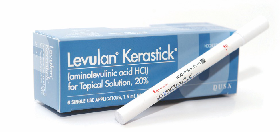
Levulan® Kerastick® (aminolevulinic acid HCl) for Topical Solution, 20% (Levulan Kerastick) plus blue light illumination using the BLU-U® Blue Light Photodynamic Therapy Illuminator (Levulan PDT) is indicated for the treatment of minimally to moderately thick actinic keratosis of the face or scalp. Actinic keratoses (AKs) are rough-textured, dry, scaly patches on the skin that can lead to skin cancer. It is important to treat AKs because there is no way to tell when or which lesions will progress to squamous cell carcinoma (SCC), the second most common form of skin cancer. So, now’s the time to manage your damage!

Levulan PDT, a 2-part treatment, is unique because it uses a light activated drug therapy to destroy AKs. How does it work? Levulan Kerastick Topical Solution is applied to the AK. The solution is then absorbed by the AK cells where it is converted to a chemical that makes the cells extremely sensitive to light. When the AK cells are exposed to the BLU-U Blue Light Illuminator, a reaction occurs which destroys the AK cells.
The 2-part treatment offers the following conveniences:
- No prescription to fill
- No daily medication to remember
- Treatment is administered by a qualified healthcare professional
Levulan PDT can also fit your lifestyle:
- The 2-part, 2 office visit treatment is completed in less than 24 hours
- Low downtime*
- High ratings for cosmetic response
- No scarring reported to date
*Patients treated with Levulan PDT should avoid exposure of the photosensitized lesions to sunlight or prolonged or intense light for at least 40 hours.


